| CAS NO: | 1158838-45-9 |
| 规格: | ≥98% |
| 包装 | 价格(元) |
| 5mg | 电议 |
| 25mg | 电议 |
| 50mg | 电议 |
| 100mg | 电议 |
| 250mg | 电议 |
| 500mg | 电议 |
| Molecular Weight (MW) | 588.07 |
|---|---|
| Formula | C31H31ClFN7O2 |
| CAS No. | 1158838-45-9 |
| Storage | -20℃ for 3 years in powder form |
| -80℃ for 2 years in solvent | |
| Solubility (In vitro) | DMSO: 118 mg/mL (200.6 mM) |
| Water: <1 mg/mL | |
| Ethanol: <1 mg/mL | |
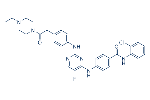
| Other info | Chemical Name: N-(2-Chlorophenyl)-4-[[2-[[4-[2-(4-ethyl-1-piperazinyl)-2-oxoethyl]phenyl]amino]-5-fluoro-4-pyrimidinyl]amino]-benzamide InChi Key: AKSIZPIFQAYJGF-UHFFFAOYSA-N InChi Code: InChI=1S/C31H31ClFN7O2/c1-2-39-15-17-40(18-16-39)28(41)19-21-7-11-24(12-8-21)36-31-34-20-26(33)29(38-31)35-23-13-9-22(10-14-23)30(42)37-27-6-4-3-5-25(27)32/h3-14,20H,2,15-19H2,1H3,(H,37,42)(H2,34,35,36,38) SMILES Code: O=C(NC1=CC=CC=C1Cl)C2=CC=C(NC3=NC(NC4=CC=C(CC(N5CCN(CC)CC5)=O)C=C4)=NC=C3F)C=C2 |
| Synonyms | TC S 7010; TCS 7010; TC-S7010 |
| In Vitro | In vitro activity: Aurora A Inhibitor I is a 2,4-dianilinopyrimidine that selectively and potently inhibits Aurora A. Aurora A Inhibitor I effectively inhibits the proliferation of HCT116 and HT29 cells, with IC50 of 190 nM and 2.9 μM, respectively. The Aurora A selectivity of Aurora A Inhibitor I against Aurora B depends on a single amino acid (Thr217) of Aurora A. In KCL-22 cells, Aurora A Inhibitor I (1-5 μM) increases G2/M cell fraction, induces histone H3 serine 10 phosphorylation, and suppresses mitotic Aurora A autophosphorylation on Thr288. Aurora A Inhibitor I (0.5-5 μM) also suppresses cell proliferation in KCL-22 cells, as well as BCR-ABL-negative leukemia cell lines KG-1 and HL-60. Aurora A Inhibitor I effectively induces apoptosis in KCL-22 cells at 5 μM. In a recent study, Aurora A Inhibitor I is also found to inhibit cell growth of HCT116, HT29, and HeLa cells, with IC50 of 377.6 nM, 5.6 μM, and 416 nM. Kinase Assay: Both Auroras A and B are assayed in ELISA format using a GST fusion (pGEX-4T) of the N-terminus of Histone H3 (aa 1–18) as substrate. Plates are coated with 2 μg/mL substrate in PBS then blocked with 1 mg/mL I-block in PBS. Kinase reactions are run for 40 min with 5 ng/mL (0.16 nM) Aurora A or 45 ng/mL (1.1 nM) Aurora B at 30 μM ATP (~ Km) in kinase buffer. Final DMSO concentration is 4%. Product is detected by incubation with antiphosphohistone H3 (Ser10) 6G3 mouse monoclonal antibody and sheep-anti-mouse HRP conjugate, followed by washing and addition of TMB substrate. After quenching with 1 M phosphoric acid, plates are read at 450 nM. Cell Assay: Cells (HCT116 and HT29 cells) are seeded in 384-well plates on day 0 in 50 μL of complete medium and incubated overnight in a 5% CO2atmosphere at 37 °C. On day 1, 10 μL of Aurora A Inhibitor I is added. On day 4, plates are allowed to reach room temperature, and 30 μL Cell Titer-Glo reagent is added to each well to measure total ATP levels. Plates are read after shaking 15 min at room temperature. |
|---|---|
| In Vivo | NA |
| Animal model | NA |
| Formulation & Dosage | NA |
| References | J Med Chem. 2009 May 28;52(10):3300-7; Carcinogenesis. 2012 Feb;33(2):285-93; ACS Chem Biol. 2012 Jan 20;7(1):185-96. |
 m.cnreagent.com