| CAS NO: | 347174-05-4 |
| 规格: | ≥98% |
| 包装 | 价格(元) |
| 1mg | 电议 |
| 2mg | 电议 |
| 5mg | 电议 |
| 10mg | 电议 |
| 25mg | 电议 |
| 50mg | 电议 |
| 100mg | 电议 |
| 250mg | 电议 |
| 500mg | 电议 |
| Molecular Weight (MW) | 262.35 |
|---|---|
| Formula | C15H22N2O2 |
| CAS No. | 347174-05-4 |
| Storage | -20℃ for 3 years in powder form |
| -80℃ for 2 years in solvent | |
| Solubility (In vitro) | DMSO: 52 mg/mL (198.2 mM) |
| Water: <1 mg/mL | |
| Ethanol: 52 mg/mL (198.2 mM) | |
| Solubility (In vivo) | 2% DMSO+50% PEG 300+5% Tween 80+ddH2O: 5mg/mL |
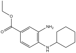
| SMILES Code | O=C(OCC)C1=CC=C(NC2CCCCC2)C(N)=C1 |
| Synonym | Frer-1; 3-amino-4-(cyclohexylamino)-benzoic acid, ethyl ester; Ferrostatin-1; Chemical Name: 3-amino-4-(cyclohexylamino)-benzoic acid, ethyl ester |
| In Vitro | In vitro activity: Ferrostatin-1 (2 μM) prevents erastin-induced ferroptosis in cancer cells, as well as glutamate-induced cell death in postnatal rat brain slices. Ferrostatin-1 is a lipid ROS scavenger, with the N-cyclohexyl moiety serving as a lipo-philic anchor within biological membranes. Ferrostatin-1 does not inhibit extracellular signal -regulated kinase (ERK) phos-phorylation or arrest the proliferation of HT-1080 cells, suggesting that it does not inhibit the MEK/ERK pathway, chelate iron, or inhibit protein synthesis. Ferrostatin-1 does, however, prevent erastin-induced accumulation of cytosolic and lipid ROS. Ferrostatin-1 readily oxidizes the stable radical 2,2-diphenyl-1-picrylhydrazyl (DPPH) under cell-free conditions. Kinase Assay: The cells treated for 24 h with Ferrostatin-1 are washed three times with phosphate-buffered saline (PBS) and pelleted by centrifugation. The supernatant is removed and 80 μL of lysis buffer is added to the cells and then stored overnight at -20°C. Subsequently, the cells are centrifuged at 10,000 RPM for 12 min and both the pellet and supernatant are stored for future use. Cell Assay: BJ-TERT/LT/ST/RASV12 cells are seeded in 100 mm dishes and allowed to grow overnight. Cells are treated with erastin (5 or 10 μg/ml) for 6, 8, or 11 hr. A camptothecin-treated (0.4 μg/ml) control is maintained, treated at the time of seeding for 20 hours. After the treatment, cells are harvested with trypsin/EDTA and washed once with fresh medium containing serum and then twice with phosphate-buffered saline. Cells are resuspended in 1× binding buffer. 100 μL is incubated with 5 μL of Annexin V-FITC and propidium iodiode for 15 min in the dark at room temperature. Then 400 μl of the 1× binding buffer s added and the cells analyzed by flow cytometry. Data are acquired and analyzed using Cellquest software. Only viable cells that do not stain with propidium iodiode are analzyed for Annexin V-FITC staining using the FL1 channel. |
|---|---|
| In Vivo | |
| Animal model | |
| Formulation & Dosage | |
| References | Cell. 2012 May 25;149(5):1060-72; Protein J. 2015 Oct;34(5):349-58. |
 m.cnreagent.com